Description
Pinealon is a synthetic linear tripeptide (Glu-Asp-Arg) with free amino and carboxyl termini; its two acidic residues and one basic arginine make it very hydrophilic. It contains no methionine, cysteine or tryptophan, the residues most prone to oxidation; as with other lyophilised peptides, keep vials sealed, cold and away from light.
Pinealon: what the published research shows
Compiled by NADA-Labs from the peer-reviewed literature cited below. No own study data.
Pinealon (Glu-Asp-Arg), also called EDR peptide, is a synthetic tripeptide studied mainly as a ‘peptide bioregulator’. Despite its name, the literature describes it as a synthetic derivative of Cortexin, a polypeptide complex from calf cerebral cortex, rather than as a pineal-gland peptide (Kuznik 2019, PMID 31099784). The research literature attributes its effects mainly to restriction of reactive oxygen species (ROS) accumulation and necrotic cell death under oxidative stress, preservation of dendritic spines in models of Alzheimer’s disease, promotion of dendrite growth (more primary dendrites, greater total length) in a model of neuronal ageing, and modulation of brain caspase-3 activity; a direct gene-regulatory action through DNA binding has been proposed on the basis of docking and biophysical data. It was studied in rodent and human-derived cell cultures, rat models of prenatal hyperhomocysteinaemia, hypoxia and carotid artery occlusion, transgenic Alzheimer’s model mice and a few Russian-language human reports.
Evidence base: Mainly in vitro and rodent in vivo studies; six of the eight numbered studies include authors from the St. Petersburg Institute of Bioregulation and Gerontology (Khavinson group), and five are summarised from abstracts without variance or p-values (three Russian-language articles). Human data: one comparative study of pinealon and vesugen (Lys-Glu-Asp, KED) in 32 adults with organic brain syndrome, without described control group, randomisation or blinding8, and three Russian-language reports without group size or on pinealon combined with vesugen (Myakotnykh 2016, PMID 28539017; Nazimko 2012, PMID 22708445; Bashkireva 2012, PMID 23734521). No randomised placebo-controlled trial, pharmacokinetic study or formal toxicology study was identified.
Key studies at a glance
| Study | Model | Design | Main result |
|---|---|---|---|
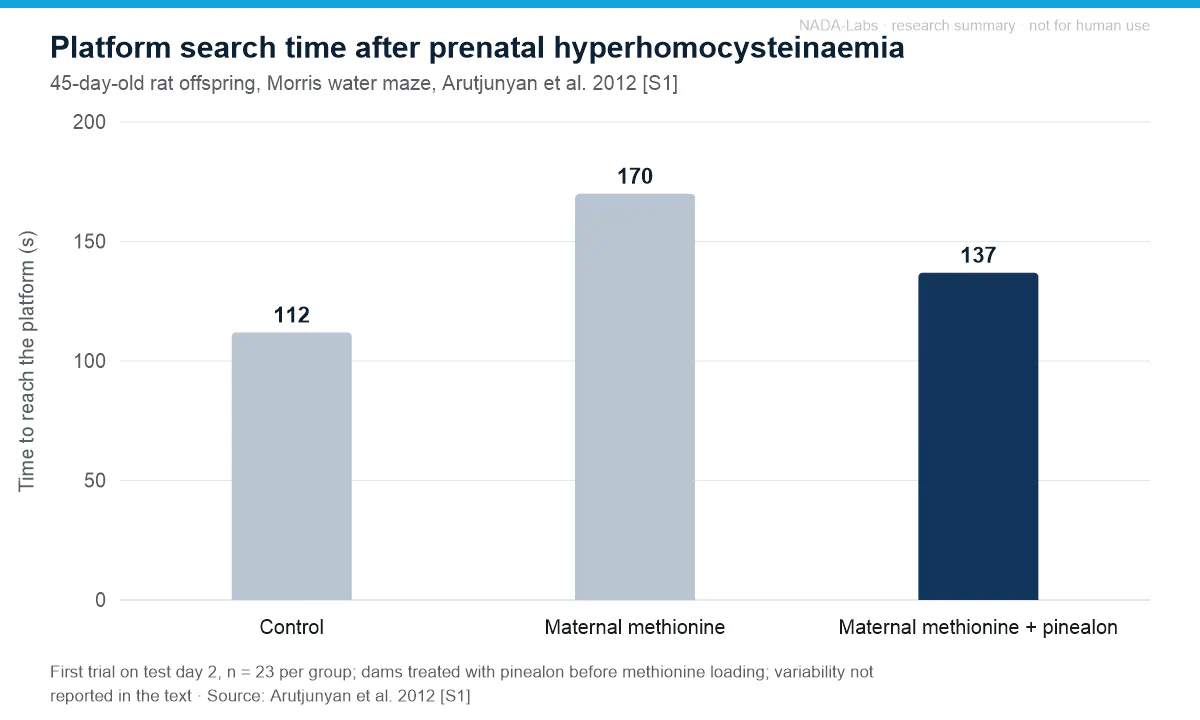
| Arutjunyan 20121 | Rat offspring after maternal methionine loading, in vivo / ex vivo | Control, methionine, methionine + pinealon (dams pretreated, intraperitoneal); n = 23/group | Maze latency 170 s (methionine) vs. 137 s (pinealon group; control 112 s); necrotic neurons after H2O2 17.7 % vs. 7.0 %; homocysteine not lowered |
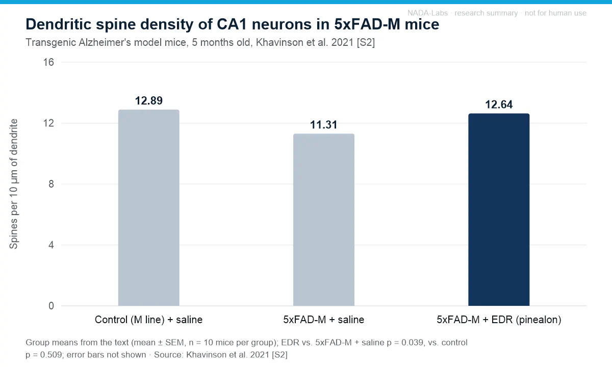
| Khavinson 20212 | 5xFAD and 5xFAD-M mice (Alzheimer’s model), in vivo | Saline vs. EDR or KED (intraperitoneal, 2 months); n = 10/group; docking | CA1 spine density 12.64 (EDR) vs. 11.31 (saline) spines/10 µm (+11 %, p = 0.039; M-line control 12.89); LTP trend not significant; predicted promoter-binding motifs |
| Mendzheritskiĭ 20113 | Old rats, carotid artery occlusion, in vivo | Pinealon vs. cortexin before occlusion; sham-operated controls | Higher survival with both; with pinealon reduced orientation, motivation and motor activity and moderately higher brain caspase-3; no effect sizes in abstract |
| Mendzheritski 20134 | Young and old rats, acute hypoxic hypoxia, in vivo | Pinealon vs. cortexin; water maze; caspase-3 in cortex and brainstem | Better maze learning than with cortexin at both ages; regional caspase-3 changes; no effect sizes in abstract |
| Khavinson 20115 | Cerebellar granule cells, neutrophils, PC12 cells, in vitro | Oxidative stress with or without pinealon; concentration series | Less ROS accumulation (concentration-dependent) and necrotic death, delayed ERK1/2 activation, cell-cycle changes; no effect sizes in abstract |
| Kraskovskaya 20176 | Mouse hippocampal neurons, amyloid synaptotoxicity, in vitro | EDR or KED vs. amyloid alone | Mushroom spines +71 % with EDR (normal level restored), +20 % with KED; n and variance not given in abstract |
| Kraskovskaya 20247 | Induced neurons from fibroblasts of 3 elderly female donors, in vitro | EDR, KED or AEDG (Ala-Glu-Asp-Gly), 10 days, vs. untreated | EDR: primary dendrites +28 % (p = 0.0208), dendrite length +46 % (p = 0.0332); 8-OHdG -23 % (p = 0.0566, not significant) |
| Meshchaninov 20158 | Human, 32 adults (41-83 years) with organic brain syndrome | Pinealon vs. vesugen; control, randomisation and blinding not described | Changes in biological-age indicators reported (larger with vesugen); pro-oxidant activity; fewer CD34+ cells; no effect sizes in abstract |
Offspring outcomes after prenatal hyperhomocysteinaemia (Arutjunyan 2012 [S1])
| Outcome (mean ± SD) | Control | Methionine | Methionine + pinealon (dams pretreated) | Statistics (as reported) |
|---|---|---|---|---|
| Body weight, age 10 days (g; 6 litters/group) | 23.05 ± 1.45 | 17.86 ± 3.05 | 25.03 ± 4.86 | methionine vs. control and pinealon vs. methionine p < 0.05 |
| Plasma total homocysteine, age 45 days (µM) | 5.6 ± 0.6 (n = 5) | 9.8 ± 0.2 (n = 4) | 10.5 ± 0.2 (n = 4) | both methionine groups vs. control p < 0.05 |
| Water-maze latency, age 45 days, trial 1 of test day 2 (s; n = 23/group; no SD in text) | 112 | 170 | 137 | methionine vs. control p < 0.01; pinealon vs. methionine and vs. control p < 0.05 |
| Cerebellar neurons (age 10 days): ROS after H2O2 (DCF, arbitrary units) | not reported | 158.7 ± 3.8 | 108.6 ± 4.1 | pinealon vs. methionine p < 0.05 |
| Cerebellar neurons: necrotic (PI-positive) cells after H2O2 (%) | not reported | 17.7 ± 2.1 | 7.0 ± 0.9 | pinealon vs. methionine p < 0.05 |
Effects reported in the studies
- Oxidative stress and cell survival in vitro. In cerebellar granule cells, neutrophils and PC12 cells under oxidative stress, pinealon restricted ROS accumulation concentration-dependently and reduced necrotic cell death, with delayed ERK1/2 activation and cell-cycle changes; the authors conclude a direct interaction with the genome (no effect sizes in abstract)5.
- Prenatal hyperhomocysteinaemia model (rat offspring). Offspring of dams pretreated with pinealon before methionine loading needed 137 s to reach the water-maze platform in trial 1 of test day 2, versus 170 s without the peptide and 112 s in controls (p < 0.05 vs. both); by trial 5 all groups performed alike. Cerebellar neurons of 10-day-old pups from this group showed fewer necrotic cells after H2O2 (7.0 % vs. 17.7 %) and less ROS (both p < 0.05), while plasma homocysteine stayed elevated; the authors attribute the effect to reduced homocysteine neurotoxicity, not lower blood levels1.
- Dendritic spines in Alzheimer’s disease models. In mouse hippocampal neurons under amyloid synaptotoxicity, EDR increased mushroom spines by 71 % to the normal level, compared with 20 % for KED (no n or variance in abstract)6. In 5-month-old 5xFAD-M mice, CA1 spine density was 12.64 spines per 10 µm with EDR versus 11.31 with saline (+11 %, p = 0.039) and not significantly different from saline-treated control M-line mice (12.89; p = 0.509); in 5xFAD mice, LTP showed only a non-significant positive trend2.
- Ageing human-derived neurons in vitro. In cortical neurons induced from fibroblasts of three elderly female donors, EDR for 10 days increased primary dendrites by 28 % (p = 0.0208) and total dendrite length by 46 % (p = 0.0332); changes in branching points (+65 %), terminal processes (+51 %) and the oxidative DNA-damage marker 8-OHdG (-23 %, p = 0.0566) were not significant7.
- Hypoxia and ischaemia models in rats. Under acute hypoxic hypoxia, pinealon improved water-maze learning in young and old rats more than the cortex-derived preparation cortexin, and the peptides changed caspase-3 activity and content region-specifically in cortex and brainstem4; given before carotid artery occlusion in old rats, both peptides increased survival (abstracts without effect sizes)3.
- Proposed gene-regulatory mechanism (in vitro and in silico). FITC-labelled pinealon entered the nucleus of HeLa cells (Fedoreyeva 2011, PMID 22117547); biophysical data indicate partial penetration of EDR into the DNA major groove (Silanteva 2019, PMID 30762356); docking predicted EDR binding motifs in promoters of genes such as CASP3, NES, GAP43 and APOE, with likely low selectivity, and the authors call the exact mechanism unknown2.
- Human reports. In 32 adults aged 41-83 years with polymorbidity and organic brain syndrome in remission, the authors report changes in biological-age indicators that they interpret as slower ageing, more pronounced with vesugen than with pinealon (no effect sizes, control group or randomisation reported)8.
Adverse effects and tolerability reported
- Behavioural changes and caspase-3 activation in old rats. With pinealon before carotid artery occlusion, orientation, motivational behaviour and motor performance decreased, and brain caspase-3 activity rose moderately in sham-operated and occluded rats3.
- Pro-oxidant and haematological findings in humans. In the 32-participant study, chemiluminescence showed pro-oxidant activity and blood CD34+ haematopoietic cells decreased, interpreted by the authors as inhibition of haemopoiesis; the abstract assigns these findings to neither peptide, mentions no clinical adverse events and bases its safety statement only on unchanged chromatin condensation8.
- Raised intracellular ROS level in vitro. Four short peptides including pinealon showed no direct antioxidant activity in vitro and raised the stationary intracellular ROS level; all except epithalon, including pinealon, at the same time lowered the share of dead neurons (Kozina 2008, PMID 18546826).
- No impairment of cultured neurons reported. At the concentration studied the peptides did not impair the morphology of human-derived induced neurons, and mitochondrial and lysosomal activity and p16 did not change significantly7.
- Missing safety and pharmacokinetic data. No formal toxicology or pharmacokinetic study was identified; blood-brain barrier passage has only been hypothesised2 and transporter uptake only predicted by docking (Khavinson 2023, PMID 36979488).
Limitations of the evidence. Most data come from one research network around the St. Petersburg Institute of Bioregulation and Gerontology, without independent replication of the in vivo findings. The mushroom-spine results of the 5xFAD-M study contradict each other, so only spine density is reported here2, and the induced-neuron study’s abstract presents a non-significant reduction of oxidative DNA damage (p = 0.0566) as an effect7. In the hyperhomocysteinaemia model the dams, not the offspring, received the peptide, and the flow-cytometry data lack a control without methionine1. The few human reports are Russian-language and qualitative, and the main study gives an inconsistent participant count (32 stated, 18 men and 12 women listed)8. None of the findings summarised here establish efficacy or safety for any use outside the described research models.
References
- Arutjunyan A, Kozina L, Stvolinskiy S, Bulygina Y, Mashkina A, Khavinson V (2012). Pinealon protects the rat offspring from prenatal hyperhomocysteinemia. International Journal of Clinical and Experimental Medicine. PMID 22567179
- Khavinson V, Ilina A, Kraskovskaya N, Linkova N, Kolchina N, Mironova E, Erofeev A, Petukhov M (2021). Neuroprotective Effects of Tripeptides-Epigenetic Regulators in Mouse Model of Alzheimer’s Disease. Pharmaceuticals. PMID 34071923 DOI
- Mendzheritskiĭ AM, Karantysh GV, Ivonina KO (2011). Effects of introduction of short peptides before carotid artery occlusion on behaviour and caspase-3 activity in the brain of old rats [article in Russian]. Advances in Gerontology = Uspekhi Gerontologii. PMID 21809624
- Mendzheritski AM, Karantysh GV, Abramchuk VA, Ryzhak GA (2013). Effect of peptide geroprotectors on the navigation system learning and caspase-3 in brain structures in rats of different age [article in Russian]. Advances in Gerontology = Uspekhi Gerontologii. PMID 28976148
- Khavinson V, Ribakova Y, Kulebiakin K, Vladychenskaya E, Kozina L, Arutjunyan A, Boldyrev A (2011). Pinealon increases cell viability by suppression of free radical levels and activating proliferative processes. Rejuvenation Research. PMID 21978084 DOI
- Kraskovskaya NA, Kukanova EO, Lin’kova NS, Popugaeva EA, Khavinson VK (2017). Tripeptides Restore the Number of Neuronal Spines under Conditions of In Vitro Modeled Alzheimer’s Disease. Bulletin of Experimental Biology and Medicine. PMID 28853087 DOI
- Kraskovskaya N, Linkova N, Sakhenberg E, Krieger D, Polyakova V, Medvedev D, Krasichkov A, Khotin M, Ryzhak G (2024). Short Peptides Protect Fibroblast-Derived Induced Neurons from Age-Related Changes. International Journal of Molecular Sciences. PMID 39518916 DOI
- Meshchaninov VN, Tkachenko EL, Zharkov SV, Gavrilov IV, Katyreva IuE (2015). Effect of synthetic peptides on aging of patients with chronic polymorbidity and organic brain syndrome of the central nervous system in remission [article in Russian]. Advances in Gerontology = Uspekhi Gerontologii. PMID 26390612 human study
This section summarises findings published in the cited peer-reviewed literature for informational purposes. It is not a claim of efficacy or safety, contains no dosing or administration information by design, and does not constitute medical, veterinary or scientific advice. Results in cell cultures or animal models do not predict effects in humans.