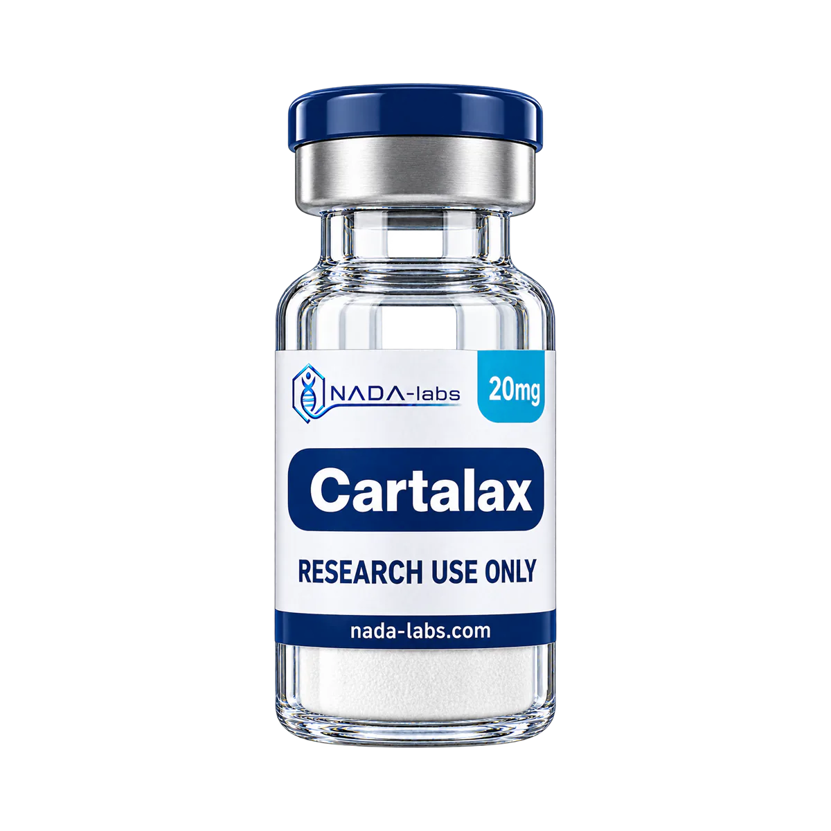
Description
Cartalax is a synthetic linear tripeptide (Ala-Glu-Asp, AED) with free amino and carboxyl termini; two of its three residues, glutamic acid and aspartic acid, carry acidic side chains. It contains no methionine, cysteine or tryptophan, the residues most prone to oxidation; as with other lyophilised peptides, keep vials sealed, cold and away from light.
Cartalax: what the published research shows
Compiled by NADA-Labs from the peer-reviewed literature cited below. No own study data.
Cartalax is the tripeptide Ala-Glu-Asp (AED), a short peptide from the ‘peptide bioregulator’ programme of Khavinson and co-workers; older papers use the code T-31, which the same group identifies as AED (Khavinson 2014, PMID 24958378). A review by the group lists AED among the short peptides of a cartilage polypeptide complex (Linkova 2023, PMID 37176122). The research literature attributes its effects mainly to a regulation of gene expression: molecular models of DNA-peptide complexes place AED in the minor groove of DNA (Khavinson 2014, PMID 25946838) and assign it to the acct sequence (Khavinson 2016, PMID 27909961), but no receptor, measured binding or pharmacokinetic study of AED was found on PubMed. It has been studied in old rats, in rats with cisplatin-induced acute renal failure and in ovariectomised rats1,2,3, and in cell cultures of human mesenchymal stem cells, skin fibroblasts and periodontal ligament stem cells4,5,6,7.
Evidence base: Three rat studies in vivo1,2,3 and four in vitro studies in human cells4,5,6,7; no clinical trial or other human study of the defined tripeptide was found on PubMed. Nearly all work comes from one research group (Khavinson, Linkova and co-authors); one cell study, written together with a second research group, declares no financial support and no conflicts of interest7. Three of the seven numbered studies are Russian-language articles available only with an English abstract1,3,6, the animal abstracts give no group sizes or variability1,2,3, and only two abstracts report numbers, partly as one range for several peptides1,4. Further cell and tissue culture work covers renal cells (Khavinson 2014, PMID 25946838; Khavinson 2014, PMID 24958378), kidney tissue explants from young and old rats (Chalisova 2015, PMID 26033601), skin fibroblasts (Lin’kova 2016, PMID 27259496), chondrocytes (Myakisheva 2023, PMID 37356100) and thymocytes (Lin’kova 2011, PMID 22238759).
Key studies at a glance
| Study | Model | Design | Main result |
|---|---|---|---|
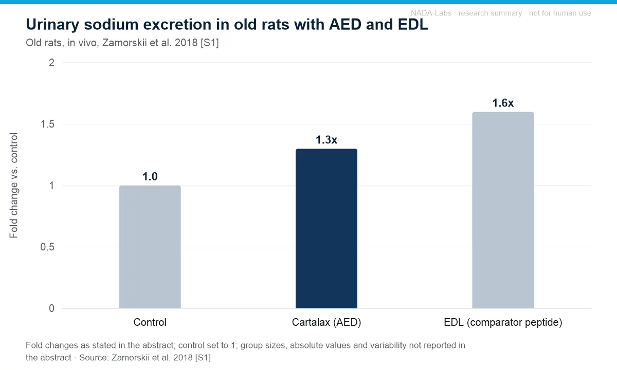
| Zamorskii 20181 | Old rats, in vivo | Comparison of a polypeptide kidney complex and the peptides AED, EDL and AEDG; renal function tests, antioxidant enzymes, peroxidation and morphology of kidney tissue; group sizes not given in the abstract | Sodium excretion 1.3x higher with AED (1.6x with EDL); diuresis 1.2-1.4x higher and urine protein 1.5-2.8x lower as ranges shared with other agents; no nephrotoxic effects according to the authors |
| Zamorskii 20152 | Rats with cisplatin-induced acute renal failure, in vivo | Comparison of a kidney polypeptide complex and the peptides AED, EDL and AEDG; renal function parameters; group sizes not given in the abstract | AED lowered urinary protein excretion and electrolyte concentration; broader normalisation of renal parameters reported for the complex, EDL and AEDG; no effect sizes in abstract |
| Povorozniuk 20073 | Ovariectomised rats (experimental osteoporosis), in vivo | Comparison of a cartilage-extract preparation and T-31 (AED); prophylactic and corrective administration; bone mineral density; group sizes not given in the abstract | Osteoprotective effect on bone mineral density reported for both substances, significantly greater with the cartilage-extract preparation; no effect sizes in abstract |
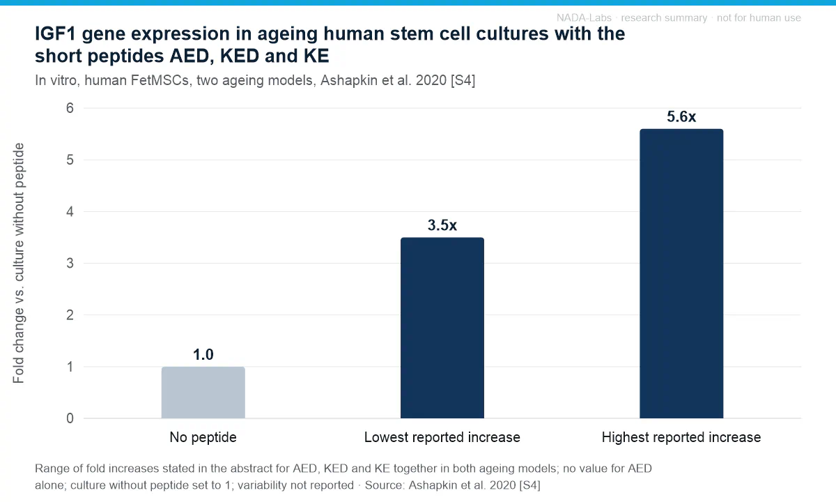
| Ashapkin 20204 | Human embryonic bone marrow mesenchymal stem cells (line FetMSCs), in vitro | Two culture ageing models (‘passages’, ‘stationary’); AED, KED and KE; expression of IGF1, FOXO1, TERT, TNKS2 and NFκB genes | IGF1 expression 3.5-5.6-fold higher with the peptides (one range for AED, KED and KE); NFκB expression raised by AED, KED and KE in both models |
| Fridman 20205 | Human skin fibroblasts during replicative ageing, in vitro | Comparison of KE and AED; immunocytochemistry and confocal microscopy of sirtuin-1, sirtuin-6, collagen I, IL-1, TGF-β and NF-κB | AED increased the synthesis of sirtuin-1, sirtuin-6 and collagen I; lower IL-1, NF-κB and TGF-β reported for KE, not AED; no effect sizes in abstract |
| Myakisheva 20236 | Human mesenchymal stem cells during replicative ageing, in vitro | Comparison of AED and a cartilage polypeptide complex; gene expression and protein synthesis of the chondrogenic differentiation markers SOX9, aggrecan, type II collagen and COMP | AED activated gene expression and protein synthesis of the markers; the complex had the same effect at a tenfold higher concentration; no effect sizes in abstract |
| Caputi 20197 | Human periodontal ligament stem cells (hPDLSCs) from 10 donors, in vitro | Six cultures (control, AEDG, KE, AED, KED, mixture); MTT proliferation assay up to 1 week; immunofluorescence (GAP43, Nestin) and western blot | GAP43 synthesis increased with AED and the other peptides on confocal microscopy; Nestin and western-blot GAP43 increases only with KED and the mixture; no inhibition of proliferation |
Renal function in old rats: reported changes for AED and the comparators (Zamorskii 2018 [S1])
| Parameter | Agents named in the abstract | Reported change |
|---|---|---|
| Diuresis | Polypeptide kidney complex, AED, EDL (one range) | 1.2-1.4x higher |
| Urine protein level and protein excretion | Polypeptide kidney complex, AED (one range) | 1.5-2.8x lower |
| Distal sodium transport | Polypeptide kidney complex, AED, EDL (one range) | 1.2-1.3x higher |
| Sodium excretion | AED / EDL | 1.3x / 1.6x higher |
| Glomerular filtration rate | AEDG only (no value for AED) | 21 % lower |
| Antioxidant enzymes and peroxidation in kidney tissue | Not attributed to individual agents in the abstract | Enzyme activity stimulated, peroxidation inhibited; together with morphology interpreted by the authors as absence of nephrotoxic effects |
Effects reported in the studies
- Renal function in old rats. AED increased urinary sodium excretion 1.3-fold (EDL 1.6-fold); for diuresis (1.2-1.4x higher) and distal sodium transport (1.2-1.3x higher) the abstract gives one range for the polypeptide kidney complex, AED and EDL together, and for urine protein level and protein excretion (1.5-2.8x lower) one range for the complex and AED1.
- Renal parameters in cisplatin-induced acute renal failure. In rats, AED decreased protein excretion and electrolyte concentration in the urine; a normalisation of diuresis, urinary creatinine, glomerular filtration rate and absolute sodium resorption was reported for the kidney polypeptide complex, EDL and AEDG, and the authors describe a potent nephroprotective effect for EDL2.
- Bone mineral density after ovariectomy. In ovariectomised rats, T-31 (AED) and a cartilage-extract preparation both showed an osteoprotective effect: they prevented the decrease of bone mineral density and increased the reduced density; the cartilage-extract preparation was significantly more effective, and the abstract gives no numbers3.
- Gene expression in ageing human mesenchymal stem cells. In FetMSC cultures aged by passaging or in stationary culture, IGF1 expression was 3.5-5.6-fold higher with the peptides — one range for AED, KED and KE — and AED, KED and KE raised NFκB expression in both models4; a later review by the same group restates the IGF1 increase for AED alone (Linkova 2023, PMID 37176122).
- Sirtuins and collagen I in ageing skin fibroblasts. In human skin fibroblasts during replicative ageing, AED increased the synthesis of sirtuin-1, sirtuin-6 and collagen I on immunocytochemistry and confocal microscopy, without effect sizes in the abstract5. In rat skin fibroblasts ageing in culture (species as given in a later review by the group, Linkova 2023, PMID 37176122), all short peptides studied, including AED, lowered MMP-9 and raised Ki-67 and CD98hc, and AED and AEDG suppressed caspase-dependent apoptosis (Lin’kova 2016, PMID 27259496).
- Chondrogenic markers in stem cells and chondrocytes. In human mesenchymal stem cells during replicative ageing, AED activated gene expression and protein synthesis of SOX9, aggrecan, type II collagen and COMP; the cartilage polypeptide complex had the same effect at a tenfold higher concentration6. In ageing chondrocytes (species not stated in the abstract), AED and the complex normalised the synthesis of molecules of the senescence-associated secretory phenotype (p16, p21, p53, TNF-α, IL-1α, Sirt1) (Myakisheva 2023, PMID 37356100).
- Neuronal differentiation marker GAP43 in periodontal ligament stem cells. In hPDLSCs from 10 donors, confocal microscopy showed more GAP43 synthesis with AED, AEDG, KE, KED and their mixture than in control cells; the increases of Nestin and of GAP43 on western blot were reported for KED and the mixture, and more than 50 % GAP43- and Nestin-positive differentiated cells for the mixture only7.
- Senescence and renewal markers in kidney cell cultures. AED and EDL increased cell proliferation, lowered the ageing markers p16, p21 and p53 and raised SIRT-6 in young and aged renal cell cultures (Khavinson 2014, PMID 25946838); in organotypic kidney tissue cultures from young and old rats, T-31 (AED) stimulated proliferation and reduced apoptosis, but to a lesser degree than the polypeptide complex (Chalisova 2015, PMID 26033601).
Adverse effects and tolerability reported
- No human study found on PubMed. The numbered studies are three rat studies and four cell-culture studies1,2,3,4,5,6,7; no clinical trial, human tolerability study, pharmacokinetic study or formal toxicology study of the defined tripeptide was found on PubMed.
- Kidney tissue in old rats. The authors interpret the stimulated antioxidant enzyme activity and lower peroxidation in kidney tissue, together with the morphological data, as absence of nephrotoxic effects; the abstract does not attribute this to individual agents and gives no mortality or body-weight data1.
- No inhibition of cell proliferation in vitro. In the MTT assay at 24, 48 and 72 h and 1 week, none of the peptides including AED inhibited the proliferation of hPDLSCs; this is a cell-culture measure, not a systemic tolerability endpoint7.
- NFκB stimulation. AED raised NFκB gene expression in both mesenchymal stem cell ageing models4; a review by the same group suggests that this effect accelerates stem cell ageing (Linkova 2023, PMID 37176122) — a suggestion by the authors, not a measured finding.
- Adverse events not reported in the abstracts. The abstracts of the kidney, bone and cell studies mention no adverse events, mortality or other safety endpoints2,3,5,6.
Limitations of the evidence. Almost all data come from one research group and its co-authors; no independent replication of the in vivo findings is among the cited records. The animal abstracts give no group sizes, variability or p-values, and several results are one range shared with other peptides or tissue complexes; in vivo, only the sodium-excretion value is given for AED alone1,2,3. Where AED was compared with the tissue polypeptide complexes, it was weaker in bone3 and in kidney tissue cultures (Chalisova 2015, PMID 26033601), and in old thymocyte cultures only another peptide (AB-9), not T-31, showed the combined effect the authors describe (Lin’kova 2011, PMID 22238759). The DNA-binding mechanism rests on docking models (Khavinson 2016, PMID 27909961); no measured binding of AED was found on PubMed. Three of the seven numbered studies are Russian-language articles with an English abstract only1,3,6, and in the stem-cell study the Nestin and western-blot increases were not seen with AED alone7. None of the findings summarised here establish efficacy or safety for any use outside the described research models.
References
- Zamorskii II, Shchudrova TS, Zeleniuk VG, Linkova NS, Nichik TE, Khavinson VK (2018). [The influence of peptides on the morphofunctional state of old rats kidneys.]. Advances in Gerontology = Uspekhi Gerontologii. PMID 30607912
- Zamorskii II, Shchudrova TS, Lin’kova NS, Nichik TE, Khavinson VKh (2015). Peptides Restore Functional State of the Kidneys During Cisplatin-Induced Acute Renal Failure. Bulletin of Experimental Biology and Medicine. PMID 26515176 DOI
- Povorozniuk VV, Khavinson VKh, Makogonchuk AV, Ryzhak GA, Kreslov EA, Gopkalova IV (2007). [Effect of peptide regulators on the structural and functional status of bone tissue in ageing rats]. Advances in Gerontology = Uspekhi Gerontologii. PMID 18306703
- Ashapkin V, Khavinson V, Shilovsky G, Linkova N, Vanuyshin B (2020). Gene expression in human mesenchymal stem cell aging cultures: modulation by short peptides. Molecular Biology Reports. PMID 32399807 DOI
- Fridman NV, Linkova NS, Kozhevnikova EO, Gutop EO, Khavinson VK (2020). Comparison of the Effects of KE and AED Peptides on Functional Activity of Human Skin Fibroblasts during Their Replicative Aging. Bulletin of Experimental Biology and Medicine. PMID 33231794 DOI
- Myakisheva SN, Linkova NS, Diatlova AS, Polyakova VO, Ryzhak GA (2023). [The influence of peptides on the chondrogenic differentiation of human mesenchymal stem cells during replicative aging.]. Advances in Gerontology = Uspekhi Gerontologii. PMID 37782646
- Caputi S, Trubiani O, Sinjari B, Trofimova S, Diomede F, Linkova N, Diatlova A, Khavinson V (2019). Effect of short peptides on neuronal differentiation of stem cells. International Journal of Immunopathology and Pharmacology. PMID 30791821 DOI
This section summarises findings published in the cited peer-reviewed literature for informational purposes. It is not a claim of efficacy or safety, contains no dosing or administration information by design, and does not constitute medical, veterinary or scientific advice. Results in cell cultures or animal models do not predict effects in humans.