Description
NAD+ is a dinucleotide coenzyme rather than a peptide, supplied here as the free acid in lyophilised form. It is markedly hygroscopic and hydrolyses in alkaline solution, so prepare stocks fresh, keep the pH near neutral and return the vial to 2–8 °C immediately after weighing.
NAD+: what the published research shows
Compiled by NADA-Labs from the peer-reviewed literature cited below. No own study data.
Nicotinamide adenine dinucleotide (NAD+) is an endogenous pyridine dinucleotide coenzyme present in every cell, where it acts as the redox carrier of glycolysis and oxidative phosphorylation and as the consumed co-substrate of sirtuin deacetylases, poly(ADP-ribose) polymerases (PARP) and CD38. The exogenous-NAD+ literature is built on the hypothesis that pathological PARP-1 activation after ischemia or trauma depletes intracellular NAD+ and that supplying the molecule directly (intranasal, intraperitoneal or intravenous route) restores tissue NAD+ and sirtuin-dependent metabolic homeostasis. Most controlled studies of NAD+ itself are rodent models of cerebral ischemia-reperfusion, traumatic brain injury, spinal cord ischemia and agonist-induced cardiac hypertrophy; the two human studies are a pharmacokinetic infusion pilot and a retrospective tolerability review. A much larger body of work concerns oral NAD+ precursors (nicotinamide riboside, nicotinamide mononucleotide), which is mentioned only for context.
Evidence base: Predominantly rat and mouse injury models (n = 6-12 per group, vehicle-, saline- or sham-controlled) plus mechanistic cell work; two of the neuroprotection papers share an author (W. Ying, UCSF). Human data on NAD+ itself are limited to one small randomized infusion pilot (n = 8 treated, 3 saline controls) and one retrospective commercial-clinic review (n = 6 NAD+, 8 NR), both with authors affiliated with clinics that provide NAD+ infusions. No randomized placebo-controlled efficacy trial of NAD+ itself was found in PubMed.
Key studies at a glance
| Study | Model | Design | Main result |
|---|---|---|---|
| Ying 20071 | Rat, transient focal cerebral ischemia | Controlled; intranasal NAD+ 2 h after onset; nicotinamide comparator | Infarct at 24/72 h profoundly reduced; neurological deficits attenuated; nicotinamide ineffective (abstract, no effect sizes) |
| Won 20122 | Rat, weight-drop traumatic brain injury | Saline-controlled; intranasal NAD+ immediately after injury; n = 6 per group | Fewer degenerating neurons in CA1, CA3, dentate gyrus (P < 0.05); cortex not protected; microglial activation in CA1 reduced at 1 and 7 d |
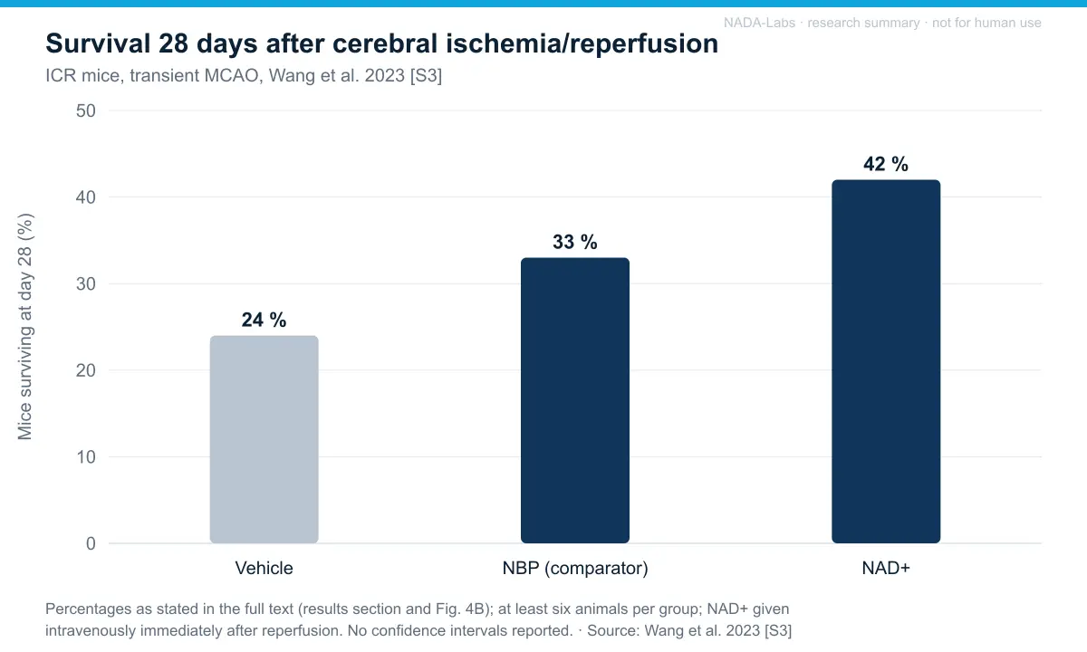
| Wang 20233 | ICR mice, transient MCAO/reperfusion; primary neurons | Vehicle-controlled; i.v. NAD+ after reperfusion; NBP comparator; n >= 6 per group; 28-day follow-up | Infarct, edema, deficits reduced at 24 h; 28-day survival 42 % vs. 24 % vehicle; ATP restored; SIRT3 up; effect abolished by sirtuin inhibitor |
| Xie 20174 | Sprague-Dawley rats, spinal cord ischemia-reperfusion | Sham, I/R, two NAD+ groups; i.p. after reperfusion; n = 12 per group; blinded BBB scoring | BBB score higher 6-24 h in higher NAD+ group (P < 0.01); apoptosis and autophagy markers reduced; lower NAD+ group no different from I/R |
| Pillai 20105 | Mice and cardiomyocytes, agonist-induced cardiac hypertrophy; SIRT3/SIRT1 genetic models | Controlled; exogenous NAD with agonist | Hypertrophic response blocked in vitro and in vivo via SIRT3-LKB1-AMPK; SIRT1-independent |
| Grant 20196 | Healthy men 30-55 y (human) | Randomized NAD+ vs. saline; continuous 6-h i.v. infusion; n = 8 + 3 | Plasma NAD+ +398 % only at 6 h; urinary NAD+ +538 %; no adverse events; liver enzymes within range |
| Reyna 20267 | Commercial-clinic clients (human) | Retrospective, no placebo; i.v. infusion series over four consecutive days, NAD+ vs. NR; 30-day follow-up; n = 6 + 8 | 6/6 NAD+ clients with moderate-severe GI and cardiovascular symptoms during infusion; infusion time 97 vs. 37 min; HDL -5.8 mg/dL; liver, renal, thyroid markers unchanged |
Human tolerability and biomarker data for intravenous NAD+
| Parameter | Grant 2019 [S6] (6-h infusion, n = 8) | Reyna 2026 [S7] (four-day infusion series, n = 6) |
|---|---|---|
| Adverse events | None observed | None documented; 6/6 with moderate-severe cramping, nausea, vomiting, tachycardia, chest pressure during infusion |
| ALT / AST | ALT unchanged; AST -3.6 U/L | No significant change at 30 d |
| ALP | Unchanged | -7.0 U/L (P = 0.04), within range |
| GGT / LD | -1.3 / -57 U/L | Not measured |
| Bilirubin | +2.75 umol/L (9.3 to 12.0, within range) | Not measured |
| HDL-cholesterol | Not measured | -5.8 mg/dL (P = 0.02) |
| HbA1c / glucose | Not measured | No change |
| hsCRP, renal, thyroid markers | Not measured | No change |
Effects reported in the studies
- Reduced ischemic brain injury (rodent stroke models). Intranasal NAD+ given 2 h after focal ischemia profoundly reduced infarct size and neurological deficits in rats, whereas nicotinamide did not (abstract, no effect sizes)1. Intravenous NAD+ immediately after reperfusion in mice reduced infarct volume, edema and deficit score at 24 h and raised 28-day survival from 24 % (vehicle) to 42 %, while restoring cortical ATP and upregulating SIRT3; protection was abolished by a sirtuin inhibitor3.
- Hippocampal neuroprotection after traumatic brain injury (rat). Intranasal NAD+ reduced Fluoro-Jade B-positive degenerating neurons in CA1, CA3 and dentate gyrus (P < 0.05, n = 6 per group) and reduced microglial activation in CA1 at 1 and 7 days, but did not protect cortical neurons or block upstream superoxide/PARP-1 activation2.
- Improved locomotor recovery after spinal cord ischemia-reperfusion (rat). In 48 rats, BBB scores were consistently higher from 6 to 24 h in the higher NAD+ group than in untreated ischemia-reperfusion (P < 0.01, n = 12 per group), with fewer TUNEL-, caspase-3- and Bax-positive neurons and reduced autophagy markers; the lower NAD+ group did not differ from untreated ischemia-reperfusion4.
- Blockade of agonist-induced cardiac hypertrophy (mouse and cardiomyocytes). Exogenous NAD maintained intracellular NAD and blocked the hypertrophic response to pro-hypertrophic agonists in vitro and in vivo via SIRT3-dependent activation of LKB1-AMPK and suppression of Akt1/mTOR signalling; the effect was absent in SIRT3 knock-out mice5. Effect sizes are not given in the abstract.
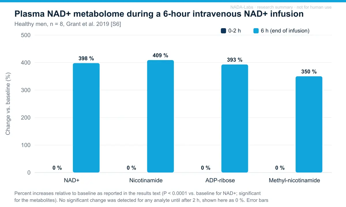
- Plasma pharmacokinetics of intravenous NAD+ (human). During a 6-h infusion in 8 healthy men, plasma NAD+ did not rise for the first 2 h and reached +398 % of baseline only at 6 h, with parallel rises in nicotinamide (+409 %), ADP-ribose (+393 %) and methyl-nicotinamide (+350 %); urinary NAD+ excretion peaked at 6 h (+538 %) and fell 43 % by 2 h after the infusion ended, consistent with rapid clearance and metabolism by NAD+ glycohydrolase/pyrophosphatase pathways6.
- Short-term biomarker changes after intravenous NAD+ (human). Over 8 h, GGT, LD and AST activities fell slightly and bilirubin rose 2.75 umol/L, all within reference ranges6. After an infusion series over four consecutive days and 30 days of follow-up (n = 6, no placebo), ALT, AST, hsCRP, renal and thyroid markers were unchanged, ALP fell 7 U/L and HDL-cholesterol fell 5.8 mg/dL; HbA1c and fasting glucose did not change7.
Adverse effects and tolerability reported
- Infusion-related symptoms. Commercial-clinic clients receiving intravenous NAD+: 6 of 6 reported moderate-to-severe abdominal cramping, diarrhea, nausea, vomiting, increased heart rate, throat pain and chest pressure during infusion, resolving immediately on completion; mean infusion time 97 min vs. 37 min for NR (P < 0.05)7. In a slow, fixed-rate 6-h infusion in 8 healthy men no adverse events were observed6.
- HDL-cholesterol decrease. -5.8 mg/dL at 30 days after the NAD+ infusion series (P = 0.02, n = 6, retrospective, no placebo); clinical relevance unclear7.
- Liver-function markers. No clinically relevant changes: GGT, LD and AST decreased and bilirubin rose slightly within reference range at 8 h6; ALT and AST unchanged and ALP -7 U/L (within range) at 30 days7.
- No toxicity reported in animal models. Intranasal NAD+ in rats described as non-toxic2; the other rodent studies do not report adverse effects1,3,4,5.
- Extracellular NAD+ as a pro-inflammatory signal (literature concern). Supraphysiological extracellular NAD+ is discussed by the authors as a danger signal with pro-inflammatory effects and as a likely driver of the infusion symptoms; not directly measured in these studies7. Grant et al. discuss extracellular catabolism of infused NAD+ by ectoenzymes CD38, CD157, CD203a and CD736.
- Tumor metabolism (precursor literature concern). NAD+ is a co-substrate of tumor-supporting pathways; in a triple-negative breast cancer mouse model, supplementation with the precursor nicotinamide riboside significantly increased cancer prevalence and metastases to the brain (Maric et al. 2023, Biosens Bioelectron, PMID 36371959; abstract, no effect sizes). No equivalent data exist for NAD+ itself.
Limitations of the evidence. Evidence for NAD+ itself rests on small rodent injury models, two of them from one research lineage, and most abstracts report significance without effect sizes; the two human datasets are pharmacokinetic and tolerability observations in 8 and 6 treated subjects, without placebo-controlled efficacy endpoints and with authors tied to clinics that provide NAD+ infusions. Infused NAD+ is cleared and metabolised rapidly, so the relationship between infusion and tissue NAD+ is unresolved. The larger precursor literature (nicotinamide riboside, nicotinamide mononucleotide) shows raised blood NAD+ in randomized trials (e.g. Martens 2018, PMID 29599478: PBMC NAD+ approx. +60 % vs. placebo, well tolerated, 24 completers of 30 randomized; Yoshino 2021, PMID 33888596: increased muscle insulin sensitivity in prediabetic women) and life-span or healthspan effects in mice (Zhang 2016, PMID 27127236; Mills 2016, PMID 28068222), but these compounds differ from NAD+ in uptake and metabolism and their results cannot be transferred to NAD+.
References
- Ying W, Wei G, Wang D, Wang Q, Tang X, Shi J, Zhang P, Lu H (2007). Intranasal administration with NAD+ profoundly decreases brain injury in a rat model of transient focal ischemia. Frontiers in Bioscience. PMID 17127275 DOI
- Won SJ, Choi BY, Yoo BH, Sohn M, Ying W, Swanson RA, Suh SW (2012). Prevention of traumatic brain injury-induced neuron death by intranasal delivery of nicotinamide adenine dinucleotide. Journal of Neurotrauma. PMID 22352983 DOI
- Wang XX, Mao GH, Li QQ, Tang J, Zhang H, Wang KL, Wang L, Ni H, Sheng R, Qin ZH (2023). Neuroprotection of NAD+ and NBP against ischemia/reperfusion brain injury is associated with restoration of sirtuin-regulated metabolic homeostasis. Frontiers in Pharmacology. PMID 37056986 DOI
- Xie L, Yu S, Wang Z, Yang K, Liu Z, Li C, Liang Y (2017). Nicotinamide adenine dinucleotide protects against spinal cord ischemia reperfusion injury-induced apoptosis by blocking autophagy. Oxidative Medicine and Cellular Longevity. PMID 28367271 DOI
- Pillai VB, Sundaresan NR, Kim G, Gupta M, Rajamohan SB, Pillai JB, Samant S, Ravindra PV, Isbatan A, Gupta MP (2010). Exogenous NAD blocks cardiac hypertrophic response via activation of the SIRT3-LKB1-AMP-activated kinase pathway. Journal of Biological Chemistry. PMID 19940131 DOI
- Grant R, Berg J, Mestayer R, Braidy N, Bennett J, Broom S, Watson J (2019). A pilot study investigating changes in the human plasma and urine NAD+ metabolome during a 6 hour intravenous infusion of NAD+. Frontiers in Aging Neuroscience. PMID 31572171 DOI human study
- Reyna K, Heinzen G, Patel N, Ritter M, Siojo A, Legere H, Pojednic R (2026). Intravenous infusion of nicotinamide adenine dinucleotide (NAD+) versus nicotinamide riboside (NR): a retrospective tolerability pilot study in a real-world setting. Frontiers in Aging. PMID 41704678 DOI human study
This section summarises findings published in the cited peer-reviewed literature for informational purposes. It is not a claim of efficacy or safety, contains no dosing or administration information by design, and does not constitute medical, veterinary or scientific advice. Results in cell cultures or animal models do not predict effects in humans.