Description
Melanotan I (afamelanotide, [Nle4, D-Phe7]-α-MSH) is a linear 13-residue analogue of α-melanocyte-stimulating hormone with an acetylated N-terminus and an amidated C-terminus, in which norleucine at position 4 and D-phenylalanine at position 7 replace methionine and L-phenylalanine. In a published forced-degradation study, the peptide broke down under acidic, basic, oxidative, UV-light and heat stress — keep vials sealed, cold and protected from light, and avoid oxidising buffers.
Melanotan I: what the published research shows
Compiled by NADA-Labs from the peer-reviewed literature cited below. No own study data.
Melanotan I (Ac-Ser-Tyr-Ser-Nle-Glu-His-D-Phe-Arg-Trp-Gly-Lys-Pro-Val-NH₂), also called afamelanotide, [Nle4, D-Phe7]-α-MSH or NDP-MSH, is a synthetic linear α-MSH analogue carrying norleucine at position 4 and D-phenylalanine at position 79 (Chawathe 2026, PMID 41547183). It was first described in 1980 as a highly potent, long-acting melanotropin resistant to serum enzymes8; in forced-degradation tests it degraded under acidic, basic, neutral, oxidative, UV-light and 60 °C stress (Chawathe 2026, PMID 41547183). The research literature attributes its effects mainly to activation of the melanocortin-1 receptor (MC1R), whose signalling increases melanin synthesis and is linked to antioxidant activity, DNA repair and modulation of inflammation (Minder 2017, PMID 28063031); in vitro the peptide also binds MC3R, MC4R and MC5R (Yang 2011, PMID 21208602), but not the ACTH receptor MC2R of mouse adrenocortical cells (Schiöth 1996, PMID 8761313). It has been studied in frog-skin and melanoma-cell bioassays, mouse models of neuroinflammation, placebo-controlled pigmentation studies in healthy volunteers, and phase 3 trials and long-term cohorts in erythropoietic protoporphyria (EPP), a photodermatosis with acute phototoxicity.
Evidence base: Mostly human data on skin pigmentation and light sensitivity: three randomised placebo-controlled volunteer studies (n = 65, 77 and 28)2,3,4, an open biopsy study (n = 7)5, two randomised, double-blind, placebo-controlled phase 3 trials in EPP (168 patients)1 and an uncontrolled long-term EPP cohort (n = 115)6. Several early studies share authors with the paper that first described the peptide4,5,8; the phase 3 trials were industry-designed and industry-sponsored1. Data outside pigmentation come mainly from animal and cell studies7. No placebo-controlled human trial outside pigmentation and photodermatoses was identified.
Key studies at a glance
| Study | Model | Design | Main result |
|---|---|---|---|
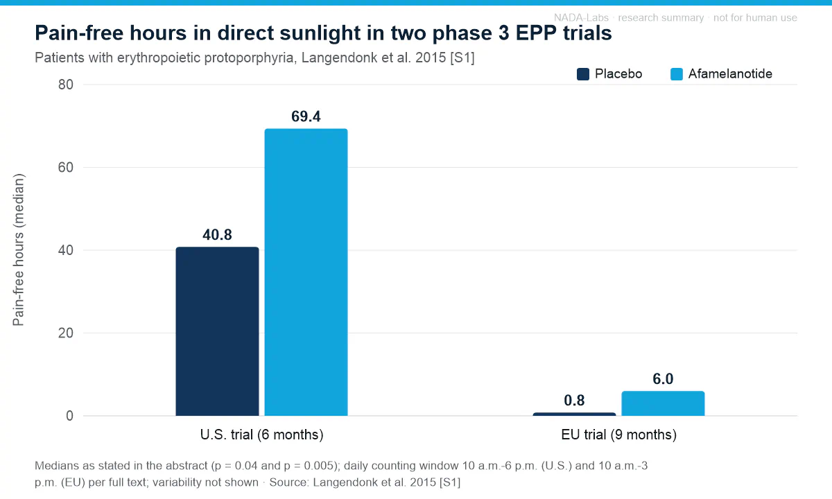
| Langendonk 20151 | Human, erythropoietic protoporphyria | Two phase 3 trials, randomised, double-blind, placebo-controlled; n = 94 (U.S., 6 months) and n = 74 (EU, 9 months) | Median pain-free time in direct sunlight 69.4 vs. 40.8 h (U.S., p = 0.04) and 6.0 vs. 0.8 h (EU, p = 0.005); disease-specific quality of life improved |
| Barnetson 20062 | Human, fair-skinned volunteers | Randomised, placebo-controlled; n = 65; 3 months | Melanin density +41 % (low minimal erythemal dose, MED) and +12 % (high MED), both p < 0.0001 vs. placebo; sunburn cells after 3 MED of UV reduced by > 50 % (low-MED volunteers); thymine dimers -59 % (p = 0.002) |
| Fitzgerald 20063 | Human, volunteers genotyped for MC1R | Randomised, placebo-controlled; n = 77 | Melanin density increased vs. placebo (p < 0.001), more in carriers of MC1R variant alleles; no effect sizes in abstract |
| Levine 19914 | Human, healthy men, skin types I-IV | Randomised, double-blind, placebo-controlled; n = 28; 7-week follow-up | Skin darkening (chromaticity) in both skin-type groups (p < 0.001), none with placebo; peak 1-3 weeks after the last administration |
| Dorr 20005 | Human volunteers, skin types III-IV | Open, uncontrolled; n = 7; skin biopsies | Eumelanin +98 % in forearm (p = 0.003) and +49 % in forehead skin (n = 3, p = 0.019); pheomelanin not significantly changed |
| Biolcati 20156 | Human, erythropoietic protoporphyria | Observational, uncontrolled; n = 115; up to 8 years | Disease-specific quality of life 31 ± 24 % of maximum before vs. 74 % after the start of treatment; only minor adverse events attributable to afamelanotide (mainly nausea) |
| Mykicki 20167 | Mouse autoimmune encephalomyelitis (EAE) in vivo; mouse and human neurons in vitro | Controlled; MC1R and orphan nuclear receptor 4 signalling | Regulatory T cells induced, EAE progression ameliorated, blood-brain barrier integrity restored, excitotoxic neuron death prevented; no effect sizes in abstract |
| Sawyer 19808 | Frog skin, mouse melanoma cells, serum, in vitro | Controlled bioassays vs. α-MSH | 26x the potency of α-MSH (adenylate cyclase); resistant to serum enzymes; prolonged activity |
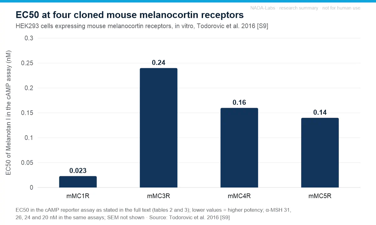
| Todorovic 20169 | HEK293 cells expressing cloned mouse melanocortin receptors, in vitro | cAMP reporter assay; alanine positional scan of α-MSH and NDP-MSH | NDP-MSH (Melanotan I) EC50 0.023 nM (mMC1R), 0.24 nM (mMC3R), 0.16 nM (mMC4R) and 0.14 nM (mMC5R); α-MSH 31, 26, 24 and 20 nM in the same assays |
Phase 3 outcomes in erythropoietic protoporphyria (Langendonk 2015 [S1])
| Endpoint | Trial | Afamelanotide | Placebo | Significance |
|---|---|---|---|---|
| Pain-free hours in direct sunlight, median (primary end point; 10 a.m.-6 p.m.) | U.S., 6 months (n = 94) | 69.4 | 40.8 | p = 0.04 |
| Pain-free hours in direct sunlight, median (primary end point; 10 a.m.-3 p.m.) | EU, 9 months (n = 74) | 6.0 | 0.8 | p = 0.005 |
| Phototoxic reactions, total number | EU, 9 months | 77 | 146 | p = 0.04 |
| Phototoxic reactions, total number | U.S., 6 months | 46 | 43 | not significant (p = 0.60) |
| Photoprovocation, hand: median change in minimum symptom dose, day 90 (J/cm²) | U.S. subgroup (n = 21) | 208.3 | 56.2 | p = 0.01 |
| Photoprovocation, lower back: same measure, day 90 (J/cm²) | U.S. subgroup (n = 21) | 227.5 | -2.4 | p < 0.001 |
Effects reported in the studies
- Potency and stability in vitro. In its first description the analogue was 26 times as potent as α-MSH in a melanoma adenylate cyclase assay, markedly more active in frog-skin and tyrosinase assays, longer acting and resistant to serum enzymes8.
- Skin darkening and eumelanin in volunteers. In 28 healthy men (randomised, double-blind), chromaticity measurements showed skin darkening in both skin-type groups (p < 0.001) and none on placebo, peaking 1-3 weeks after the last administration4. In an open study in seven volunteers, biopsy eumelanin rose one week after the last administration by 98 % in the forearm (p = 0.003) and 49 % in the forehead (n = 3, p = 0.019), with no significant change in forearm pheomelanin5.
- Melanin density and UV-damage markers. In 65 fair-skinned volunteers, melanin density measured by reflectance spectroscopy rose on average by 41 % in the low-MED skin type (2.55 -> 3.59) and by 12 % in the high-MED type (4.18 -> 4.70; both p < 0.0001 vs. placebo); after UV exposure at three times the minimal erythemal dose, epidermal sunburn cells were reduced by more than 50 % in low-MED volunteers and thymine dimers in the basal layer by 59 % (p = 0.002)2. In 77 genotyped volunteers the increase in melanin density versus placebo (p < 0.001) was greater in carriers of six MC1R variant alleles than in non-carriers3.
- Light tolerance in erythropoietic protoporphyria. In both phase 3 trials median pain-free time in direct sunlight was longer with afamelanotide than with placebo (69.4 vs. 40.8 h, U.S.; 6.0 vs. 0.8 h, EU; different daily counting windows), and in a U.S. subgroup the light dose provoking symptoms rose more than with placebo. Phototoxic reactions were fewer only in the EU trial; the disease-specific EPP-QOL score improved, whereas the Dermatology Life Quality Index did not change1.
- MC1R signalling beyond pigmentation (mouse, in vitro). In mice with EAE, the peptide (as NDP-MSH) induced functional regulatory T cells, ameliorated disease progression and restored blood-brain barrier integrity; in mouse and human neurons in vitro it prevented excitotoxic death, a neuroprotection mediated via MC1R and orphan nuclear receptor 47.
Adverse effects and tolerability reported
- Tolerability in the phase 3 trials. Adverse events were generally mild to moderate, most commonly headache, nausea, nasopharyngitis and back pain; clinically relevant group differences were limited to mild implant-site hyperpigmentation in one third of afamelanotide recipients and darkened moles in a few. Serious adverse events (four on afamelanotide, including a benign compound naevus; two on placebo, including a melanoma) were considered unrelated to the study drug1.
- Volunteer and long-term studies. Three small phase 1 studies reported nausea and transient facial flushing as minor toxic effects and no pathological findings at UV-exposed skin sites (Dorr 2004, PMID 15262693); the long-term EPP study recorded only minor adverse events attributable to the peptide, mainly nausea6.
- Immunogenicity. No anti-drug antibodies were detected in 23 of 26 patients exposed for up to 6 years; three patients had pre-existing immunoreactivity whose titres did not change (Lengweiler 2015, PMID 25402764).
- Melanocytic lesions. Case reports after unregulated use of melanotan I and II describe changes in existing moles and new (dysplastic) naevi; four case reports describe melanomas arising from existing moles during or shortly after melanotan use; conclusive evidence of a causal link is lacking (Habbema 2017, PMID 28266027).
- Haemodynamic effects in mice. In conscious mice, NDP-α-MSH raised blood pressure and heart rate dose-dependently, with a more prominent tachycardic than pressor effect; the MC3/4 receptor antagonist SHU9119 abolished these effects (Rinne 2012, PMID 22982611).
Limitations of the evidence. The placebo-controlled volunteer studies are small (n = 28-77), two of them without effect sizes in the abstract3,4; the biopsy eumelanin data are uncontrolled (n = 7)5. The two phase 3 trials differed in end points, duration and number of study-drug administrations, were partially unblinded by the increased skin pigmentation and were industry-sponsored1; the long-term data are uncontrolled6, and effects outside pigmentation rest on animal and cell studies7. Human results were obtained with clinical trial preparations under medical supervision and refer to those trial populations, not to this research material. None of the findings summarised here establish efficacy or safety for any use outside the described research models.
References
- Langendonk JG, Balwani M, Anderson KE, Bonkovsky HL, Anstey AV, Bissell DM, Bloomer J, Edwards C, Neumann NJ, Parker C, Phillips JD, Lim HW, Hamzavi I, Deybach JC, Kauppinen R, Rhodes LE, Frank J, Murphy GM, Karstens FPJ, Sijbrands EJG, de Rooij FWM, Lebwohl M, Naik H, Goding CR, Wilson JHP, Desnick RJ (2015). Afamelanotide for Erythropoietic Protoporphyria. New England Journal of Medicine. PMID 26132941 DOI human study
- Barnetson RS, Ooi TK, Zhuang L, Halliday GM, Reid CM, Walker PC, Humphrey SM, Kleinig MJ (2006). [Nle4-D-Phe7]-alpha-melanocyte-stimulating hormone significantly increased pigmentation and decreased UV damage in fair-skinned Caucasian volunteers. Journal of Investigative Dermatology. PMID 16763547 DOI human study
- Fitzgerald LM, Fryer JL, Dwyer T, Humphrey SM (2006). Effect of MELANOTAN, [Nle(4), D-Phe(7)]-alpha-MSH, on melanin synthesis in humans with MC1R variant alleles. Peptides. PMID 16293341 DOI human study
- Levine N, Sheftel SN, Eytan T, Dorr RT, Hadley ME, Weinrach JC, Ertl GA, Toth K, McGee DL, Hruby VJ (1991). Induction of skin tanning by subcutaneous administration of a potent synthetic melanotropin. JAMA. PMID 1658407 DOI human study
- Dorr RT, Dvorakova K, Brooks C, Lines R, Levine N, Schram K, Miketova P, Hruby V, Alberts DS (2000). Increased eumelanin expression and tanning is induced by a superpotent melanotropin [Nle4-D-Phe7]-alpha-MSH in humans. Photochemistry and Photobiology. PMID 11045725 DOI human study
- Biolcati G, Marchesini E, Sorge F, Barbieri L, Schneider-Yin X, Minder EI (2015). Long-term observational study of afamelanotide in 115 patients with erythropoietic protoporphyria. British Journal of Dermatology. PMID 25494545 DOI human study
- Mykicki N, Herrmann AM, Schwab N, Deenen R, Sparwasser T, Limmer A, Wachsmuth L, Klotz L, Köhrer K, Faber C, Wiendl H, Luger TA, Meuth SG, Loser K (2016). Melanocortin-1 receptor activation is neuroprotective in mouse models of neuroinflammatory disease. Science Translational Medicine. PMID 27797962 DOI
- Sawyer TK, Sanfilippo PJ, Hruby VJ, Engel MH, Heward CB, Burnett JB, Hadley ME (1980). 4-Norleucine, 7-D-phenylalanine-alpha-melanocyte-stimulating hormone: a highly potent alpha-melanotropin with ultralong biological activity. Proceedings of the National Academy of Sciences of the United States of America. PMID 6777774 DOI
- Todorovic A, Ericson MD, Palusak RD, Sorensen NB, Wood MS, Xiang Z, Haskell-Luevano C (2016). Comparative Functional Alanine Positional Scanning of the α-Melanocyte Stimulating Hormone and NDP-Melanocyte Stimulating Hormone Demonstrates Differential Structure-Activity Relationships at the Mouse Melanocortin Receptors. ACS Chemical Neuroscience. PMID 27135265 DOI
This section summarises findings published in the cited peer-reviewed literature for informational purposes. It is not a claim of efficacy or safety, contains no dosing or administration information by design, and does not constitute medical, veterinary or scientific advice. Results in cell cultures or animal models do not predict effects in humans.